|
Bioss
bs 6694r  Bs 6694r, supplied by Bioss, used in various techniques. Bioz Stars score: 91/100, based on 1 PubMed citations. ZERO BIAS - scores, article reviews, protocol conditions and more https://www.bioz.com/result/bs 6694r/product/Bioss Average 91 stars, based on 1 article reviews
bs 6694r - by Bioz Stars,
2026-04
91/100 stars
|
Buy from Supplier |
|
Bio-Techne corporation
ap-2 gamma antibody Ap 2 Gamma Antibody, supplied by Bio-Techne corporation, used in various techniques. Bioz Stars score: 90/100, based on 1 PubMed citations. ZERO BIAS - scores, article reviews, protocol conditions and more https://www.bioz.com/result/ap-2 gamma antibody/product/Bio-Techne corporation Average 90 stars, based on 1 article reviews
ap-2 gamma antibody - by Bioz Stars,
2026-04
90/100 stars
|
Buy from Supplier |
|
R&D Systems
ap2 gamma Ap2 Gamma, supplied by R&D Systems, used in various techniques. Bioz Stars score: 94/100, based on 1 PubMed citations. ZERO BIAS - scores, article reviews, protocol conditions and more https://www.bioz.com/result/ap2 gamma/product/R&D Systems Average 94 stars, based on 1 article reviews
ap2 gamma - by Bioz Stars,
2026-04
94/100 stars
|
Buy from Supplier |
|
R&D Systems Hematology
goat monoclonal tfap2c 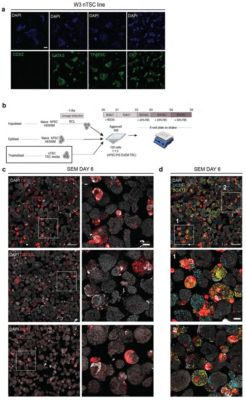 Goat Monoclonal Tfap2c, supplied by R&D Systems Hematology, used in various techniques. Bioz Stars score: 93/100, based on 1 PubMed citations. ZERO BIAS - scores, article reviews, protocol conditions and more https://www.bioz.com/result/goat monoclonal tfap2c/product/R&D Systems Hematology Average 93 stars, based on 1 article reviews
goat monoclonal tfap2c - by Bioz Stars,
2026-04
93/100 stars
|
Buy from Supplier |
|
Proteintech
anti tfap2c Anti Tfap2c, supplied by Proteintech, used in various techniques. Bioz Stars score: 93/100, based on 1 PubMed citations. ZERO BIAS - scores, article reviews, protocol conditions and more https://www.bioz.com/result/anti tfap2c/product/Proteintech Average 93 stars, based on 1 article reviews
anti tfap2c - by Bioz Stars,
2026-04
93/100 stars
|
Buy from Supplier |
|
Novus Biologicals
anti ap 2 Anti Ap 2, supplied by Novus Biologicals, used in various techniques. Bioz Stars score: 90/100, based on 1 PubMed citations. ZERO BIAS - scores, article reviews, protocol conditions and more https://www.bioz.com/result/anti ap 2/product/Novus Biologicals Average 90 stars, based on 1 article reviews
anti ap 2 - by Bioz Stars,
2026-04
90/100 stars
|
Buy from Supplier |
|
Proteintech
anti g2 cop antibody Anti G2 Cop Antibody, supplied by Proteintech, used in various techniques. Bioz Stars score: 85/100, based on 1 PubMed citations. ZERO BIAS - scores, article reviews, protocol conditions and more https://www.bioz.com/result/anti g2 cop antibody/product/Proteintech Average 85 stars, based on 1 article reviews
anti g2 cop antibody - by Bioz Stars,
2026-04
85/100 stars
|
Buy from Supplier |
|
The AP 2 gamma Antibody AP2g 6E4 4 Biotin from Novus Biologicals is a mouse monoclonal antibody to AP 2 gamma This antibody reacts with human mouse The AP 2 gamma Antibody AP2g 6E4 4
|
Buy from Supplier |
|
The AP 2 gamma Antibody AP2g 6E4 4 DyLight 550 from Novus Biologicals is a mouse monoclonal antibody to AP 2 gamma This antibody reacts with human mouse The AP 2 gamma Antibody AP2g 6E4
|
Buy from Supplier |
|
Sequence-specific DNA-binding protein that interacts with inducible viral and cellular enhancer elements to regulate transcription of selected genes. AP-2 factors bind to the consensus sequence 5'-GCCNNNGGC-3' and activate genes involved in a large spectrum of
|
Buy from Supplier |
|
Sequence-specific DNA-binding protein that interacts with inducible viral and cellular enhancer elements to regulate transcription of selected genes. AP-2 factors bind to the consensus sequence 5'-GCCNNNGGC-3' and activate genes involved in a large spectrum of
|
Buy from Supplier |
Image Search Results
Journal: Frontiers in Cell and Developmental Biology
Article Title: ESRRB Facilitates the Conversion of Trophoblast-Like Stem Cells From Induced Pluripotent Stem Cells by Directly Regulating CDX2
doi: 10.3389/fcell.2021.712224
Figure Lengend Snippet: Antibodies used in this experiment.
Article Snippet: CDX2 ,
Techniques:
Journal: bioRxiv
Article Title: Transgene-Free Ex Utero Derivation of A Human Post-Implantation Embryo Model Solely from Genetically Unmodified Naïve PSCs
doi: 10.1101/2023.06.14.544922
Figure Lengend Snippet: a, immunofluorescence images validating correct expression of TSC marker genes in colonies of a WIBR3 (W3) naïve hESC derived TSC line (termed nTSC). CK7, TFAP2C, GATA3, CDX2 (all in green); DAPI (blue). Scale bar, 100 µm. b, scheme for aggregation protocol of naïve pluripotent stem cells (nPSCs) in HENSM media, naïve-derived trophoblast stem cells (nTSCs), and nPSCs induced in RCL towards PrE/ExEM for 3 days. c , immunofluorescence images showing rare expression of CK7, GATA3, and SDC1 (red) trophoblast markers in the aggregates. nuclei (DAPI, white); Left column scale bar, 500 µm; Right, zoom into several aggregates with CK7 expression; scale bar, 100 µm. d , immunofluorescence image from (upper left panel in c) , showing Epi (OCT4, cyan) and PrE (SOX17, yellow) with CK7 (red); nuclei (DAPI, white). Zoom ins from the indicated regions are shown. Although some aggregates express lineage markers, they do not organize into embryoid-like structures and are not uniformly surrounded by the trophoblast compartment. Scale bar, 500 µm; bottom, 100 µm.
Article Snippet: The antibodies and dilutions employed for cell immunofluorescence were the following: Rabbit polyclonal anti-Cdx2 (Cell Signaling Cat# 3977) 1:200; Mouse monoclonal anti-Cdx2 (Biogenex Cat# MU392A-UC) 1:200; Rabbit polyclonal anti Gata4 (Abcam Cat# Ab84593) 1:120; Rabbit monoclonal anti-Foxa2 (Abcam Cat# Ab108422) 1:100; Goat polyclonal anti-Sox17 (R&D Cat# AF1924) 1:200; Mouse monoclonal anti-Oct4 (clone C-10) (Santa Cruz Cat# SC-5279) 1:200; Rabbit monoclonal Cdx2 (Abcam Cat# ab76541) 1:200;
Techniques: Immunofluorescence, Expressing, Marker, Derivative Assay

Journal: bioRxiv
Article Title: Transgene-Free Ex Utero Derivation of A Human Post-Implantation Embryo Model Solely from Genetically Unmodified Naïve PSCs
doi: 10.1101/2023.06.14.544922
Figure Lengend Snippet: a, scheme of the donor plasmid vector used for genomic integration of the DOX-inducible iGATA3 overexpression transgene. b , immunofluorescence images of iGATA3 cells, showing uniform GATA3 expression (green) only in response to DOX; nuclei (DAPI, blue). Scale bars, 100 µm. c , brightfield images of WT (top) and iGATA3 (bottom) cells after incubation in BAP(J) media for three days. Scale bar, 200 µm. d , qRT-PCR gene expression (normalized by GAPDH and ACTIN) of the trophoblast markers for WT naïve pluripotent stem cells (nPSCs) in BAP(J) media (purple) and iGATA3 nPSC cells induced by DOX in BAP(J) media (yellow), versus nPSCs maintained in HENSM media and used as a reference control (set as 1) (white). e , immunofluorescence images showing different patterns of expression of trophoblast marker genes in the wild type (WT) nPSCs incubated in BAP(J) media for three days (top) versus iGATA3 cells, induced by DOX in BAP(J) media (bottom). GATA3 (magenta), TFAP2C (magenta), GATA2, CDX2, SDC1, and HCGB (all in green), nuclei (DAPI, blue). Scale bar, 100 µm. f , immunofluorescence images showing uniform expression of CK7 in colonies of WT nPSCs incubated in BAP(J) media for three days (top), whereas colonies of iGATA3 cells induced by DOX in BAP(J) media express CK7 heterogeneously, with most of them being negative for CK7. Scale bar, 100 µm. g , from left to right, FACS plots of ENPEP versus TACSTD2 for trophoblast (Tb) priming using iGATA3 induction in different media (AP(J) with and without DOX, BAP(J) with and without DOX), and using WT nPSC priming to trophectoderm in AP(J) and BAP(J) regimens. Percentage of double positive population is indicated. This result shows that transient expression of the GATA3 transgene is not required for Tb differentiation and that it can be achieved with BAP(J) media using WT nPSCs as starting cells.
Article Snippet: The antibodies and dilutions employed for cell immunofluorescence were the following: Rabbit polyclonal anti-Cdx2 (Cell Signaling Cat# 3977) 1:200; Mouse monoclonal anti-Cdx2 (Biogenex Cat# MU392A-UC) 1:200; Rabbit polyclonal anti Gata4 (Abcam Cat# Ab84593) 1:120; Rabbit monoclonal anti-Foxa2 (Abcam Cat# Ab108422) 1:100; Goat polyclonal anti-Sox17 (R&D Cat# AF1924) 1:200; Mouse monoclonal anti-Oct4 (clone C-10) (Santa Cruz Cat# SC-5279) 1:200; Rabbit monoclonal Cdx2 (Abcam Cat# ab76541) 1:200;
Techniques: Plasmid Preparation, Over Expression, Immunofluorescence, Expressing, Incubation, Quantitative RT-PCR, Gene Expression, Control, Marker